
Spinal Compression Fracture
The Causes Behind Spinal Compression Fracture

By Neurosurgery Singapore
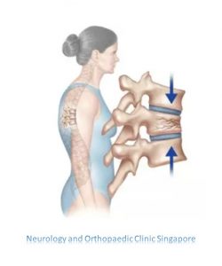
What is Spinal Compression Fracture?
A spinal compression fracture caused by osteoporosis is commonly referred to as a compression fracture, but it can also be called a vertebral, osteoporotic, or wedge fracture. It occurs when a vertebral bone in the spine loses at least 15-20% of its height due to fracture. While wedge fractures are the most common type, other types of compression fractures can also occur, such as:
- Crush fracture. If the entire bone breaks, rather than just the front of the vertebra, it may be called a crush fracture.
- Burst fracture. This type of fracture involves a loss of height in both the front and back walls of the vertebral body, rather than just the front. This distinction is important because burst fractures can be unstable, potentially leading to progressive deformity or neurological complications.
What are some symptoms of Spinal Compression Fracture?
Along with back pain, spinal compression fractures also can cause:
- Pain that gets worse when you stand or walk but with some relief when you lie down
- Trouble bending or twisting your body
- Loss of height
- A curved, stooped shape to your spine
The pain typically happens with a slight back strain during an everyday activity like:
- Lifting a bag of groceries
- Bending to the floor to pick something up
- Slipping on a rug or making a misstep
- Lifting a suitcase out of the trunk of a car
- Lifting the corner of a mattress when changing bed linens
“Fractures caused by osteoporosis most often occur in the spine. Spinal fractures — called vertebral compression fractures — occur an estimated 1.5 million times each year in the United States. “
We have a solution to stop the pain. Click here to let us help you.
What types of diagnosis?
To diagnose compression fracture, our Specialist will take a complete medical history and perform a physical examination. If a compression fracture is suspected, our doctor may order the following diagnostic procedures:
- X-ray (also known as plain films) –use invisible electromagnetic energy beams to produce images of bones. While soft tissue structures like the spinal cord, nerves, discs, and ligaments are not visible, X-rays provide a detailed view of bone anatomy, spinal alignment, and curvature. They can assess conditions like spinal dislocation (spondylolisthesis), kyphosis, and scoliosis, as well as spine balance. Specific bone abnormalities, such as bone spurs, disc space narrowing, fractures, or erosion, can also be detected. Dynamic X-rays, such as flexion/extension images, can show spine movement and help identify any abnormal motion or instability at the affected levels.
- Magnetic resonance imaging (MRI) – a diagnostic procedure that uses strong magnets, radio waves, and computer technology to create detailed images of organs and structures inside the body.
- Computed tomography scan (CT scan): a diagnostic imaging procedure that uses X-rays and computer technology to create detailed images of the body, including bones, muscles, fat, and organs. CT scans provide more detailed views than standard X-rays, allowing for a clearer examination of various body structures.
- Nuclear bone scan: a diagnostic procedure in which a small amount of radioactive material is injected into the body to measure bone activity. The radiation used is minimal—less than that in half of a CT scan. This scan is useful in detecting damaged or abnormal bones.
Since compression fractures usually occur in weakened bone, our doctor may order additional tests to look for osteoporosis, tumor, or infection.



Possible treatment methods?
Treatment for a compression fracture depends on the nature of the break and the likely cause. Our Specialist non-surgical approaches include:
Taking pain medications
Resting in bed until better
Wearing a back brace
Going to physical therapy
Treating osteoporosis if bone weakening is suspected
If the body does not respond well enough to recovery, the surgical options include:
Fusion: Repairing the bone and joining vertebrae together after an injury
Tumor treatment: Removing bone (if needed) to treat a tumor
Kyphoplasty: The procedure involves using a small balloon, inserted through a needle, to raise the vertebrae and then filling the space with special cement. This treatment can help relieve pain from a vertebral fracture that persists for more than six weeks, though it carries certain risks. It’s important to discuss these potential risks with our doctor.
